-5973916109880.jpg)
THE FIRST TRAINING COURSE LASTED FOR 2 DAYS OF AUGUST 26-27, 2022 AT THE HCM CITY IN THE SOUTHERN PROVINCES OF VIETNAM
NEUROLOGICAL DISEASES PROGRAM
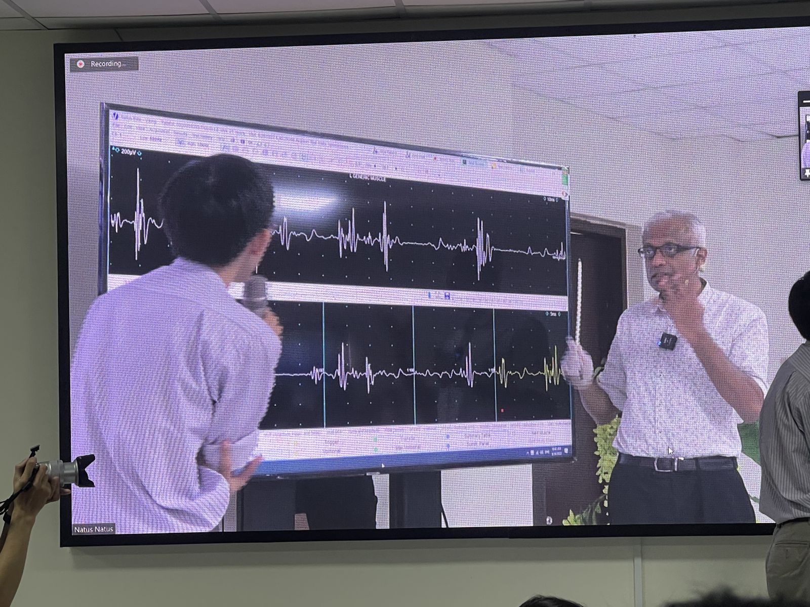
ELECTRODIAGNOSIS OF BRACHIAL PLEXOPATHY
A/Prof T. Umapathi
Senior Consultant, Department of Neurology
National Neuroscience Institute, Singapore

Table 1: Common causes of brachial plexopathy
|
Pathologic categories |
Specific causes |
|
Inflammatory |
Brachial Neuritis (Parsonage –Turner syndrome) Multifocal motor neuropathy Radiation plexitis |
|
Neoplastic |
Metastatic breast carcinoma |
|
Trauma |
Motorcycle accidents Fall from height Backpack injuries Obstetric Erb’s palsy Post-CABG |
|
Congenital/ hereditary |
Hereditary neuropathy with liability for pressure palsies (HNPP) Hereditary Neuralgic Amyotrophy True neurogenic thoracic outlet syndrome |
There are four main aims to an electrodiagnostic study of the brachial plexus:
1) Localise pathology to the brachial plexus
2) Map accurately the parts of the plexus that have borne the brunt of the injury. This must be done in “two planes”, i.e. proximal (roots) vs. distal (cords/nerves); as well as upper, middle and lower sections of the brachial plexus.
Objectives 1 & 2 require a good understanding of:
• Functional anatomy of the brachial plexus
• More importantly, the differential utility of various upper limb electrodiagnostic studies to help accurately delineate the lesion within the brachial plexus.
3) Assess if pathology is predominantly demyelinating or axonal.
This helps decide the etiology of the brachial plexopathy and prognosticate for neurological recovery. For instance, demyelination points to neuropraxic trauma or inflammatory causes like multifocal motor neuropathy. Conversely, axonopathy would indicate axontemesis or neurontemesis in traumatic plexopathies.
4) Estimate severity of neural injury, and therefore again help
prognosticate recovery.
Aim 1: Localise pathology to the brachial plexus.
Understaning the functional anatomy of the
brachial plexus.
Exercise 1
C5
C6
C7
C8
T1
Brachial plexus--- a simple approach to its anatomy
C5, C6, C7, C8 T1 form the brachial plexus.
C5, C6 make upper trunk
C8, T1 make lower trunk
C7 continues as the middle trunk
Alltrunks must divide into anterior and posterior divisions.
All 3 posterior divisions form the posterior cord which becomes the radial nerve after the axillary nerve exits.
The anterior division of the lower trunk becomes the medial cord, which then becomes the ulnar nerve after the medial cutaneous nerve of the forearm and branch to median leave.
The remaining anterior divisions of upper and middle trunks make lateral cord. The lateral cord leads to musculocutaneous nerve and gives a branch to median nerve.
The musculocutaneous nerve becomes the lateral cutaneous nerve of the forearm after giving a branch to biceps.
Finally add the suprascapular nerve to the upper trunk ---**
Table 2: The path of individual nerves within the brachial plexus.
|
Nerve |
AHC/roots |
Trunk |
Division |
Cord |
Termination |
|
Axillary |
C5,6 |
Upper |
Posterior |
Posterior |
Deltoid , teres minor |
|
Musculo Cutaneous |
C5,6 |
Upper |
Anterior |
Lateral |
Biceps, Lat cut nerve of forearm (C6) |
|
Radial |
C6, 7, 8 |
Middle Upper Lower |
Posterior |
Posterior |
EI, digit I (C7,8) |
|
Ulnar |
C8, T1 |
Lower |
Anterior |
Medial |
ADM, 1st DIO, digit V (C8) |
|
Median |
C6, 7, 8,T1 |
Upper |
Anterior |
Lateral |
Digits I, II PT, FCR |
|
Middle |
Digits III, II PT, FCR |
||||
|
Lower |
Medial |
APB |
|||
|
Medial cut nerve of the forearm |
T1 |
Lower |
Anterior |
Medial |
Medial cut nerve of the forearm |
|
Digit I, median |
C6 |
Upper |
Anterior |
Lateral |
Median |
|
Digit III |
C6 10% C7 70% C8 20% |
Middle 70% (LT 20%, UT 10%) |
Anterior |
Lateral 80% (MC 20%) |
Median |
|
Digit V |
C8 |
Lower |
Anterior |
Medial |
Ulnar |
|
Digit I, radial |
C6 60%, C7 40% |
UT 60%, MT 40% |
Posterior |
Posterior |
Radial |
Exercise 2
AIM 2: Map accurately the parts of the plexus that have borne the brunt of the injury. This must be done in “two planes”, i.e. proximal (roots) vs. distal (cords/nerves) as well as upper, middle or lower sections of the brachial plexus.
Sequence of study.
As in all electrodiagnostic studies, the initial part is a brief but focused clinical evaluation. Information to help decide on etiology, extent, severity and prognosis of plexopathy should be obtained. Attention should also be paid to the type and mechanism of trauma as it helps direct subsequent examinations.
The nerve conduction and needle electrode survey is then planned.
The first step is sensory nerve conduction studies. Depending on the findings from history and physical examination some or all of the sensory nerve studies listed in table 3 can be done. This is followed by the motor nerve conductions. Again, the studies chosen should be dictated by the earlier findings (table3).
Needle electrode examination completes the examination. It is often quite detailed necessitating survey of a number of muscles (table3).
Table 3: Nerve conduction and EMG abnormalities arising from pathology at different parts of the brachial plexus.
|
Electro Diagnostic study |
Upper trunk |
Middle trunk |
Lower trunk |
Lateral Cord |
Posterior cord |
Medial cord |
|
Sensory |
Lateral Cut. nerve of the forearm. Digit I (median) Digit I (radial 60%) |
Digit III (median) Digit I (radial 40%) |
Digit V (ulnar) Medial cut. nerve of the forearm |
Lateral cut. nerve of the forearm. Digit I, II, III (median) |
Digit I (radial) |
Digit V (ulnar). Medial cut. nerve of the forearm |
|
Motor |
Axillary nerve (deltoid) Musculocut. nerve (biceps) |
- |
Ulnar (ADM) Median (APB) Radial (EI) |
Musculocut. nerve (biceps) |
Axillary (deltoid) Radial (EI) |
Ulnar (ADM) Median (APB) |
|
EMG |
Deltoid Biceps Brachio- radialis Infra- spinatus Rhomboids Serratus anterior Mid-Cx paraspinal |
EDC ECR Brachio radialis Triceps Latorsi d Serratus anterior Mid Cx paraspinal |
APB 1St DIO EI Lower Cx paraspinal (Horner’s) |
Biceps Pronator teres FCR |
EI EDC ECR Brachio radialis Triceps Deltoid Lat dorsi |
APB 1St DIO ADM FCU FDP (IV,V) |
AIM 3 & 4: Assess pathology and severity.
Proximal brachial plexus injury, as in avulsion injury to spinal roots, carries an extremely poor prognosis. Pre-ganglionic root-level localization is suggested by the presence of normal sensory potentials (SNAPs) in the areas of sensory loss. Needle electrode examination could confirm this by finding denervation in the paraspinal muscles of the same segment.
In the acute period (at least more than a week after onset), the compound muscle action potential (CMAP) amplitude could give an idea of the severity of axon loss especially if compared with the normal contralateral side. However, this is not sensitive as up to 50% of axons may be lost before CMAP changes are noted. In chronic lesions collateral innervation from surviving axons would reduce further the utility of CMAP amplitude or area in estimating axon loss.
The SNAP amplitude is a more sensitive index of axon loss but the changes take longer, up to ten days.
At all stages, the interference pattern gives an accurate picture of lesion severity, although one has to be careful not to use this alone for prognostication. In demyelinating lesions conduction block would reduce recruitment of motor units, similar to axon loss in axonopathy. However, the prognosis for good neurological recovery is good in the former while guarded in the latter. Generally, the amount of spontaneous activity is not a good gauge of severity.
At the end of the study, the electrodiagnostician would have been able to: 3) Localize pathology to brachial plexus
4) Chart the lesion in “two planes” as elaborated above
5) One would also have also been able to assess, from the sum of nerve conduction and EMG data, whether the lesion is predominantly demyelinating or axonal and therefore help the referring doctor in deciding on the etiology (table 1). Rarely the electrodiagnostician may be able to point the clinician towards a definite cause e.g. the presence of myokymia would suggest radiation plexitis rather than compressive brachial plexopathy from metastatic breast tumour.
In summary, the key to the electro diagnostic evaluation of brachial plexus is a good understanding of its functional anatomy. The various nerve conduction and EMG examinations of the upper extremity are then utilised to accurately characterise the nature, extent and severity of the lesion.
Nerve conduction studies (NCS) – The basics of Abnormal Patterns.
In this write–up the definition of common NCS abnormalities are expanded and expounded upon. It would serve as a primer to the practical sessions where these abnormalities would be demonstrated on real patients.
CMAP- Compound muscle action potential.
A nerve trunk has thousands of axons; when stimulated extraneously by current each individual axons will activate the muscle fibers that constitutes its motor unit. The sum of the electrical activity of all the muscle fibers of a motor unit makes up its electrical potential (MOTOR UNIT POTENTIAL). In turn, a summation of the electrical activity of the many motor units within the muscles, when recorded on the surface of the muscle belly, constitutes the bell-shape electrical potential known as the COMPOUND MUSCLE ACTION POTENTIAL (CMAP). In other words, the area of the CMAP curve is made up of the sum off all the electrical potentials of individual motor units.
Some of the motor units are fast and are represented in the initial part of the CMAP. The interval from the stimulus artifact to the point of first electrical activity marks the DISTAL MOTOR LATENCY (DML). Other motor units are slow and they are at the rear end of the CMAP. The interval between the initial deflection to the point when the CMAP returns to the baseline, reflects the spread of velocities among the various motor units. This interval is the DURATION of CMAP.
The majority of motor units hover around the 50th centile, and summate to peak at this point. The voltage at this point defines the AMPLITUDE of CMAP.
Now what can go wrong?
1) Axonopathy- less axons, therefore less motor units and: ∙ CMAP area decreases,
∙ CMAP amplitude drops.
Occasionally if the axon loss involves, by chance the fastest units, a mild amount of slowing (not more than 20%) can occur.
2) Myelinopathy-all axons slow equally, therefore; ∙ CMAP DML prolongs
3) Myelinopathy- various axons slow variably, then:
∙ CMAP DML prolongs,
∙ CMAP duration prolongs as the spread of velocity difference between various motor units is increased.
As a result of this spread there is a greater chance for the peak of one motor unit potential to fall on the trough of another motor unit, “phase-cancelling” each other so that they cannot contribute to the CMAP. Therefore:
∙ CMAP amplitude drops
Conduction velocity-CV.
By stimulating two points of the nerve, at a known distance, and using the time interval between the onset of two recorded CMAPs, CONDUCTION VELOCITY-CV can be calculated.
Now what can go wrong?
1) Axonopathy-less axons:
∙ CV remains unchanged
However, if the axon loss involves, by chance, the fastest units, a mild amount of slowing (not more than 20%) can occur
2) Myelinopathy-all axons slow equally:
CV-decreases uniformly in all nerves and in all segments of the nerves, distal and proximal.
3) Myelinopathy- various axons slow variably, then:
∙ CV-decreases but not uniformly.
∙ CMAP amplitude decreases, because of the increase in phase cancellation
When recording over a longer distance of the nerve, the spread between the velocities of various motor units is further increased. This increases the phase cancellation and causes, TEMPORAL DISPERSION (TD)- Abnormal TD is defined as reduction in proximal CMAP amplitude with an increase in the duration by 30%. This occurs when the variable slowing of many axons causes phase
cancellation and a drop in CMAP amplitude; with a concomitant increase in CMAP duration. The longer the segment of nerve studied the more obvious the temporal dispersion becomes. As an analogy imagine a cohort of runners with different speeds. When they are made to run a longer, rather than a short, distance the varying speeds of individual runners would “disperse” the cohort and separate better the fastest from the slowest.
Conduction block (CB)
A reduction of the CMAP amplitude (or area) by >50% (without an increase of CMAP duration of more than 30%) between proximal and&
Đang truy cập: 6
Trong ngày: 612
Trong tháng: 5880
Tổng truy cập: 675405